Shaky

Halo
Halo
Halo/Shaky
Words properly spaced on each line
Words appear to be poorly spaced or out of position
swirl
Lines of text appear curved
Letters only in good registration in center

The abnormal conditions described below are not currently amenable to any known surgical or pharmacological intervention.
Helen Irlen, an educational psychologist working in California, has defined a series of conditions observed among her young subjects that impede their ability to read at grade. In the context of this site, these conditions are the symptoms of a variety of underlying diseases. While these diseases have been grouped under the title syndrome, they are found individually in different subjects. Due to this fact, they are labeled a pseudo-syndrome in this work.
The purpose of this webpage is to explore these symptoms from a physiological perspective in order to understand their origins.
The Irlen Institute has marketed a variety of colored filters for many years, through a staff of screeners and diagnosticians, as aids in alleviating the symptoms noted. The underlying idea has been described by Irlen (1991) and somewhat earlier by Meares (1980). It is currently not known whether the putative effect is psychological or physiological in character. Wilkins (1995) has discussed the methodology and provided references. Robinson (1998) provides statistics on similar abnormalities. The empirical results of tests found in the scientific literature (Simmers 2001) are controversial. No theoretical discussion of the effects of such filters has been located.
This webpage is drawn from Section 18.8.5.3 of the larger work Processes in Biological Vision. It will address these diseases from a physiological perspective. They can be described as electrical abnormalities within the neural system supporting vision. Most of them can be associated with errors in the oculomotor servomechanism controlling the motions of the eyes. This page calls upon many other pages of this website and other sections of the main work [by chapter and section numbers shown in brackets]. Much of this other material is not designed for the lay reader.
Irlen has caricatured the symptoms reported to her by her subjects over the years on her website. She has defined the symptoms using the descriptive names shown on the left. They have been grouped according to their apparent functional relationship as shown on the right.
| Descriptor | Cause | |
|---|---|---|
| Blurry & Washout | Imaging problems | |
| Halo and shaky | Local spatial errors | |
| Rivers, Seesaw & Swirl | Global spatial errors |
This webpage will attempt to associate more functionally and clinically explicit names with the Irlen descriptors. On initial review, the symptoms she illustrates appear to relate primarily to abnormalities in the oculomotor servomechanisms of the eyes. More specifically, to errors in the pointing of the two eyes during the reading process. Several of the caricatures represent symptoms related to various degrees of the same abnormality (disease). Two of the symptoms appear to relate to imaging errors within the perception oriented signal processing of the visual system.
Irlen has used the expression scotopic sensitivity syndrome as an alternate designation for this syndrome. This page will not use this designation. No relationship between these diseases and the term scotopic vision or the sensitivity related to scotopic vision has been uncovered in this review. Conditions that do relate to scotopic vision are addressed on a separate webpage labeled "Snowy Vision"
It is useful to present a series of test samples that attempt to capture the essence of the Irlen samples and can be used to perform a preliminary evaluation on a child exhibiting reading difficulties.
Each of the following samples can be enlarged by clicking on it. If the child is trying to describe one of these thumbnails, expand the thumbnail to full size and ask to child to look at it in an attempt to confirm the specific condition the child is attempting to describe.
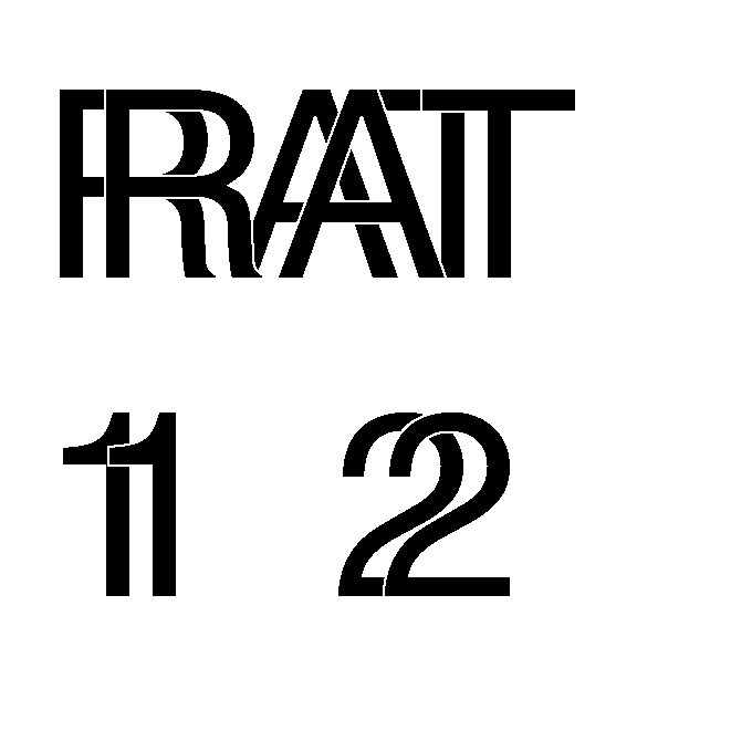
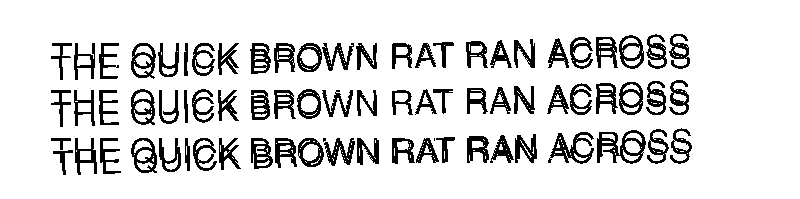
| Halo/ Shaky |
|
|
|
|||||
| Clear | Horizontal Halo | Vertical Halo | Diagonal Halo/Shaky | |||||
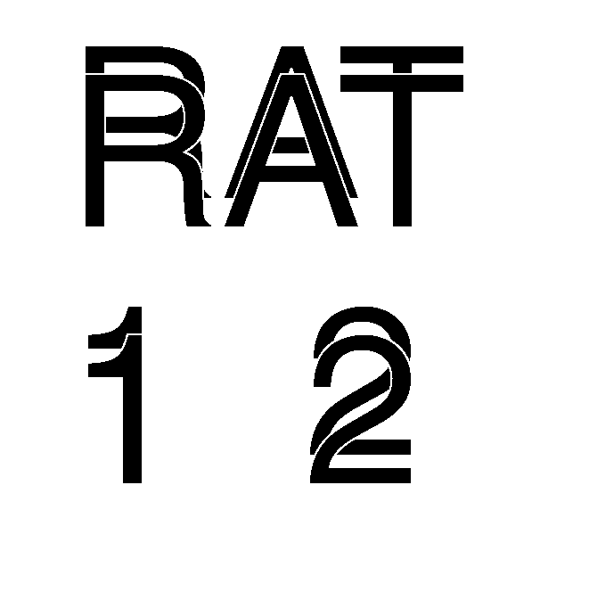
| Rivers | |
|||||||
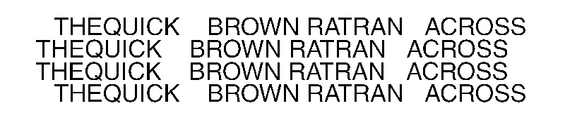
| Clear Words properly spaced on each line | Rivers Words appear to be poorly spaced or out of position | |||||||
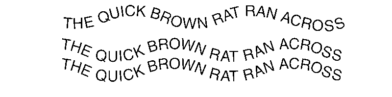
| Seesaws/ swirl |
|
|
||||||
| Seesaws Lines of text appear curved | Swirl Letters only in good registration in center | |||||||
The abnormalities associated with the above conditions can be located by using one or more of the TOP LEVEL BLOCK DIAGRAMS of Vision.
Of particular interest here is the simplified Block Diagram shown below. It related to the servomechanisms of the oculomotor system. This figure only describes one of three servomechanisms associated with the three pairs of eye muscles. It displays the horizontal servomechanism. The vertical servomechanism is similar but slightly simpler. There is also a third servomechanism that acts to control the rotation of each eyeball about its line of sight. It is considerably simpler.
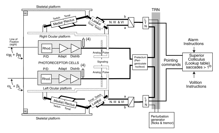
THE HORIZONTAL SERVOMECHANISM OF THE OCULOMOTOR SYSTEM
The top of the figure relates to the right eye. The bottom relates to the left eye. The superior colliculus on the right is responsible for generating the basic pointing (version) commands that cause the eyes to rotate in the same direction. These will be considered in-phase commands here. They are shown by the light signaling lines on the right.The perigeniculate nucleus (PGN, and sometimes called the pretectum)is responsible for generating the second type of pointing (vergence)commands that make the eyes converge on a point at close range. These are out-of-phase commands. They are shown by the heavy signaling lines on the right. The perturbation generator at the bottom of the picture also generate signals that are normally in-phase like those of the pointing (version) commands. The shaded area is the thalamic reticular nucleus (TRN), a structure through which all the signals pass. The TRN performs a supervisory function and can interrupt the various signals in response to alarm mode requests. It also accepts a signal from the PGN that is used to analyze fine detail projected onto the foveola of the eye.
Of particular note in this figure is the dual character of each oculomotor muscle. Each contains a tonal and a twitch element. The tonal element is used to point the eyes while the twitch element is important in the analysis of small detail (such as reading). These elements are driven by separate neural circuits in each of the servomechanisms defined above.
PRELIMINARY EXPLANATION OF SYMPTOMS/TEST SEQUENCEBy examining each of the empirically defined symptoms shown above and the preceding diagram it is possible to outline the source of the abnormal condition (disease) causing it. In summary form, with each term to be justified later, the labels provided by Irlen can be expanded as follows:
| Label | Nature of Error | Peak Error |
|---|---|---|
| Blurry | Indeterminate based on caricature | --- |
| Washout | Differentiation in the conscious signal path | (expressed in different domain) |
| Horizontal Halo | Out-of-phase error in horizontal vergence servo drive | +/-1/16 character |
| Vertical Halo | Out-of-phase error in vertical vergence servo drive | +/-1/16 character |
| Diagonal Halo/Shaky | Synchronous error in both the horizontal and vertical servo drives | +/-1/16 character |
| Rivers | In-phase error in horizontal version servo drive | one to two characters as shown |
| Seesaw | In-phase non-synchronous noise in vertical oculomotor servo. | three lines of text |
| Swirl | Error in oblique(rotation correction) oculor muscle servo drive | two degrees as shown |
This simple table highlights the need to more precisely define the magnitude of the observed error (the symptom) in terms of angular spatial coordinates. The magnitude of the symptoms are largely independent of the size of the type used in the evaluations. However, if the type size is known and the distance from the eye to the text is known, the angular parameters are easily calculated. The multiple categories in the table and the test samples also suggest the descriptor Halo might be changed to a more appropriate Twinning. In the expanded samples within the Halo category, the characters have been outlined as an aid in interpretation. However, the term shadow is not appropriate when speaking of one of the characters.
The remaining four symptoms have been illustrated by the above thumbnails. They fall into two classes.
The Halo, Shaky & Rivers conditions all appear to be mild forms of the more serious disease known as strabismus. The Seesaw condition appears to a mild form of the more serious disease known as nystagmus. The Swirl condition is suggestive of a failure in the oblique muscles of the eyes (and posssibly a failure in a neural compensation circuit). It may be useful to define (or locate in the literature) more specific scientific or medical names for these conditions.
The above material summarizes the result of the exploratory work in this area. To further pursue the detailed sources of these diseases, and quantify them calls for a next step. This step would be to design more quantitative test protocols for each of these conditions in conjunction with educational psychologists. These could be used to evaluate a statistically valid sample of those exhibiting or reporting difficulty reading.
Members of the public who find this material useful in testing their own children are encouraged to confer with their school psychologist or staff, using these pages as a basis for discusssion.
Those researchers interested in pursuing the insights provided here are encouraged to communicate with this site by Direct E-Mail
The error sources identified so far are not currently amenable to any known surgical or pharmacological intervention.
Return to the CLINICAL (Vision) Home Page .
Return to the NEURON Home Page .
Return to the website VISION Home page
References
Meares, O. (1980) Figure/background, brightness/contrast and reading disabilities Visible Language vol 14, 13-29
Irlen, H. (1991) Reading by Colours. NY: Avery
Wilkins, A. (1995) Visual Stress. NY: Oxford University Press pp 132-158
Robinson, G. (1998) The Familial Incidence of Symptoms of Scotopic Sensitivity/Irlen Syndrome http://www.newcastle.edu.au/centre/sed/irlen/paper-incidence.html
Simmers, A. Bex, P/ Smith, F. & Wilkins, A. (2001) Spatiotemporal visual function in tinted lens wearers Invest Ophthal Vis Sci vol. 42, no. 3, pp 879-884