
This webpage references sections of the manuscript, "Processes in Biological Vision" available on the web at the address shown in the references (Fulton, 2005). Section numbers of the manuscript are shown in parentheses. The first number shown is the chapter number; it is followed by the section numbers. Download individual chapters.
The subject of macular sparing has been controversial within the clinical community for a very long time. The reason has been an inadequate model of the visual system. With an expansion of the basic model, a clearer understanding of both macular sparing and foveola sparing becomes available. The difference between these two phenomena is significant both to the patient and physiologically.
This webpage will address the following subjects:
Figure 1 shows the anatomical dimensions of the retina of the right eye (viewed through the pupil) and the corresponding right eye visual field (viewed from the pupil).
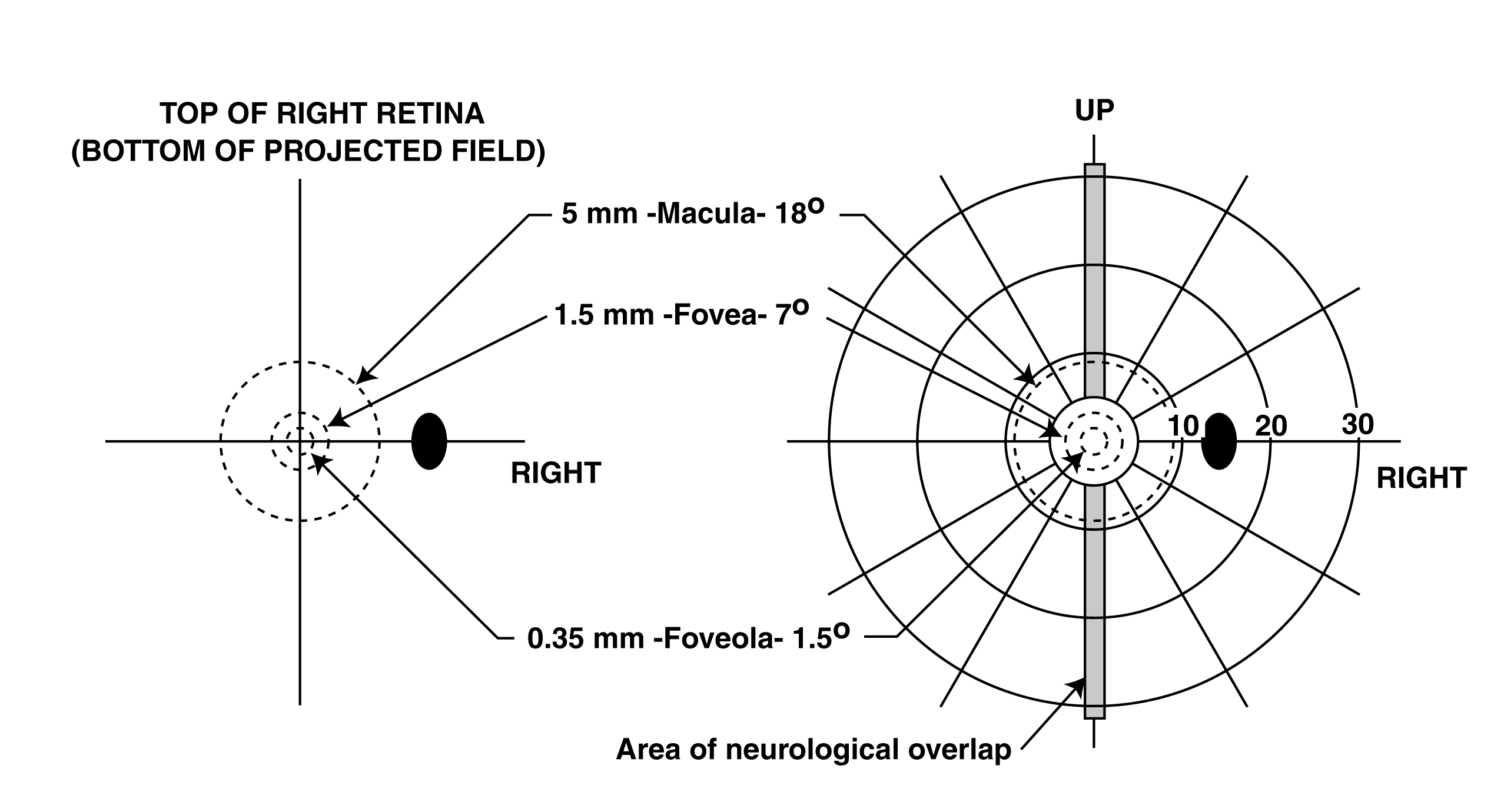
The highest resolution portion of the retina is called the foveola and it occupies an area of 0.35 mm diameter centered on the point of fixation. In the field of view, this corresponds to a diameter of 1.5 degrees.
The next highest resolution portion of the retina is called the fovea and it occupies an area surrounding the foveola and extending to a diameter of 1.5 mm (a diameter of 6.5-7 degrees in the visual field)
Both of these features are smaller than the smallest circle on the typical perimetry chart, which has a 10 degree diameter.
A highly variable diameter area usually identified by the ophthalmologist is called the macula. It is difficult to achieve consensus on the diameter of the macula because of its diffuse character. Polyak defined it as 3 mm minimum diameter (11 degrees) and 5 mm maximum diameter (18 degrees & shown here)with it extending to the edge of the papilla (optic disk or blind spot) in some preparations. The macula is not a discrete anatomical feature. It is caused by pigments diffused through the fourth through ninth layers of the retina. The value of 25 degrees used recently by Gray, Galetta & Shatz appears excessive.
The diameter of the blind spot (optical disk or papilla) is equally hard to define rigorously. It is typically described as 5 degrees in diameter horizontally and 7 degrees in diameter vertically centered 15 degrees from the point of fixation (Section 2.2.2).
There is a band of neurological overlap (typically described as extending to one degree each side of the vertical meridian, measured at the point of fixation). This band is represented in both of the hemifields projected to the occipital cortex. It will be described further below. This band is not identifiable when viewing the retina.
To understand the physiology of sparing in vision (and many other significant phenomena), it is necessary to use an expanded schematic of vision. Figure 2 shows such a schematic.
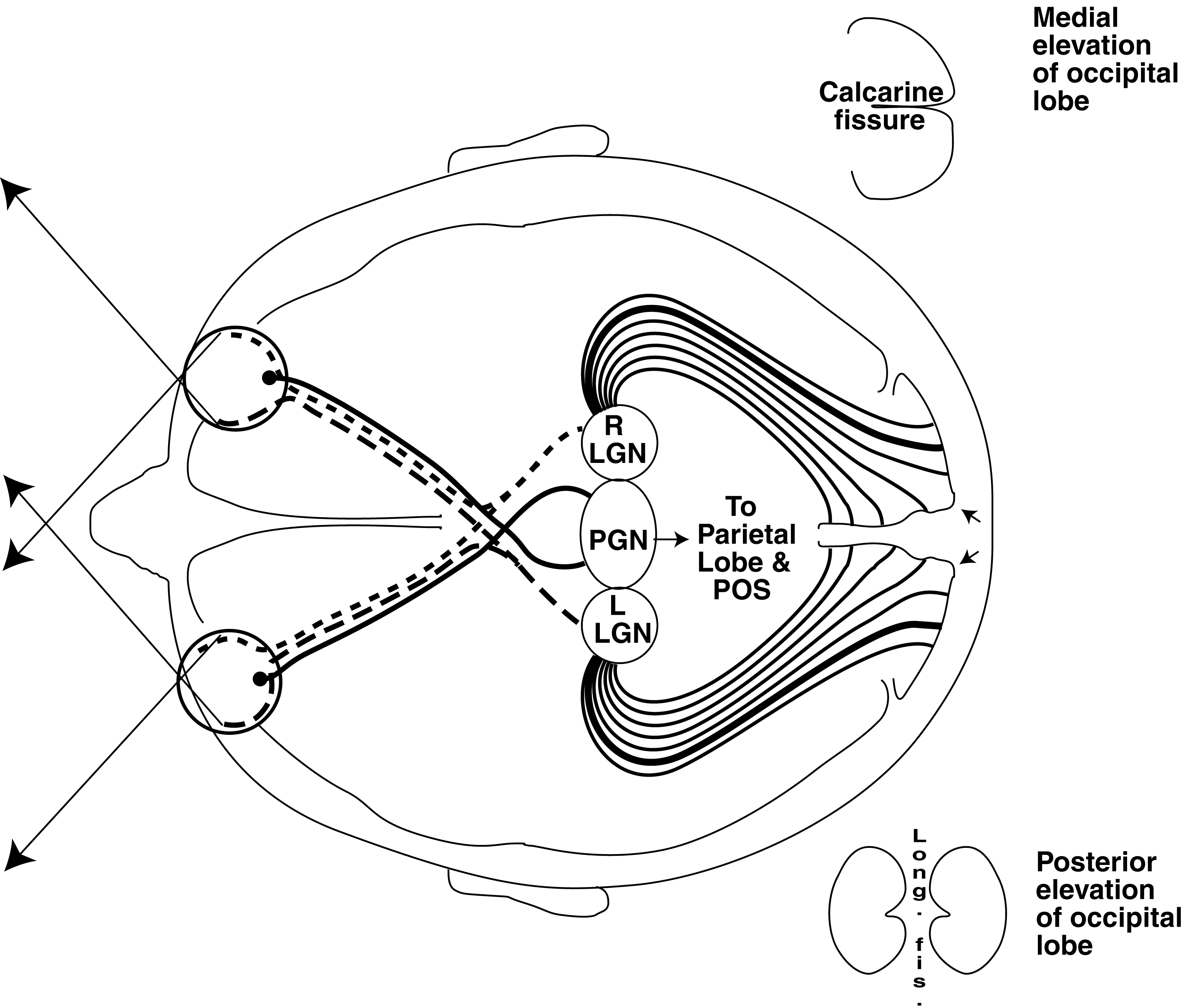
This presentation shows three signal paths from each retina. The two dashed lines represent low resolution signals from each hemifield of the retina and constitute 98% of the axons in the optic nerve. These lines merge in the optic chiasm as typically described in the literature. The solid lines represent additional signaling channels. These channels represent high resolution signals acquired from the foveola and representing one axon per photoreceptor of the foveola. The total of these axons represents only 2% of the axons in the optic nerve. Their scarcity, and the lack of an adequate model suggesting their presence, has prevented their experimental isolation and description in the past. These axons terminate at the perigeniculate nucleus (PGN) after passing through the optic chiasm (Section 15.1.6). The PGN is usually identified as a small section of the morphologically identified lateral geniculate nucleus (LGN). The functional significance of the PGN is generally greater than that of the LGN.
The introduction of an additional projection of the foveola to the PGN introduces an entirely different element to the discussion. This expanded model is compatible with both the functional evidence, the histology of the midbrain and current occipital lobe mappings.
Particularly strong evidence for the presence of the PGN is the fact that information presented to the foveola of the eyes arrives at the parietal lobe of the human brain before it arrives at the occipital lobes. The only logical path supporting this phenomenon is a direct path from the midbrain to the parietal lobe. This path is measurably shorter than the path from the midbrain to the occipital lobes and back to the parietal lobe under any conditions.
The overlay of the visual representations onto the occipital lobes is highly variable due to the folding of the cerebrum. The location of the two points of fixation are represented by the heavy lines proceeding from the LGNs to the occipital lobes. They terminate between the poles of the occipital lobes and much nearer the two ears, depending on the folding.
The blood supplies to the occipital lobes are separated into the supplies to the two walls facing the longitudinal fissure and the supplies serving the external portions of the two lobes. The blood supplies to the occipital lobes outside the longitudinal fissure are segregated into four zones separated by the longitudinal fissure and the two calcarine fissures (see posterior elevation). Details of these supplies are available in Smith & Richardson and in Liu, Volpe & Galetta.
The major failures to the neurological system are due to interruptions of the blood supply (embolisms) and lesions to and physical pressures on the neural circuits. These failures apply to all elements of the visual system. However, due to the packaging of the system, lesions due to external causes are typically restricted to the occipital lobes.
Because of the redundancies forward of the LGN/PGN, failures of the hemianopia type (or sectors of the hemifields) are typically associated primarily with the portions posterior to the LGN/PGN. Embolisms related to the various blood supplies noted above correlate well with the majority of large area anopias. These failures due to embolisms and physical factors are well documented in Neuro-ophthalmology texts, such as Liu, Volpe & Galetta.
Figure 3 illustrates three anopia situations in detail.
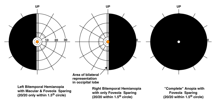
The left frame shows a Left Hemianopia with macular sparing as found following an embolism involving the right Calcarine branch of the right posterior cerebral artery (PCA). Macular sparing is observed because the right posterior temporal branch of the PCA was not involved. While not obvious, the foveola has been spared completely because of the location of the embolism. The subject will exhibit high visual acuity within the 1.5 degee field centered on the point of fixation. The subject will exhibit normal resolution within the seven degree diameter field centered on the point of fixation. Normal resolution will be observed in the area of neural overlap, except for a possible signal-to-noise loss with the loss of redundancy in this area.
The middle frame shows a Left Hemianopia with foveola sparing only. In this case, the embolism involves both the calcarine branch and the posterior temporal branch of the right PCA. There is no macular sparing but foveola sparing is observed (although a color has been added to help resolve the foveola in this frame). The subject will still exhibit high visual acuity within the 1.5 degree field centered on the point of fixation. Visual acuity in the neural overlap region will be preserved as described above.
The right frame shows complete anopia with foveola sparing (also known as keyhole vision, tunnel vision or bitemporal hemianopia). This situation is to be expected in the case of bilateral embolisms or lesions affecting both lobes of the occipital region. The keyhole has a diameter of about 175 photoreceptors with about 23,000 photoreceptors within its area. This is sufficient photoreceptor area to read about 11 or 12 characters at a comfortable type size. This size illustrates why reading involves a salutatory process consisting of reading short words or syllables individually in sequence (Chapter 19). Keyhole vision has been studied most completely by Huber. He noted foveola sparing was a characteristic of all his case studies and they all achieved normal visual acuity through this keyhole. He also noted there was no sign of bifurcation of the foveola in any of his cases.
Many other more complex, and less symmetrical, forms of anopia are documented in the literature.
The clinical community has long debated whether the point of fixation is projected to and represented in both lobes of the occipital (rear-most) section of the brain. The arguement has centered on whether the projections from the two eyes were unilateral or bilateral. The actual situation cannot be described using these simple terms alone. Recent experiments have demonstrated the fact the projections to the occipital lobes are unilateral except for a narrow vertical field of about one degree that is projected to both lobes of the occipital region. Even this extension of the discussion does not satisfy all of the functional evidence.
The clinical community has also struggled with the recognized fact of "blind sight," at least perception of objects in the visual field in the absence of functional occipital lobes. This blind sight generally takes two forms.
The question of unilateral versus bilateral representation was further obscured by the fact that the region they associated with the foveola was not bifurcated along the vertical meridian like the outer regions of the visual fields. The above discussion demonstrates that each full foveola is represented at high resolution in the PGN. The two images are merged within the PGN prior to information extraction. At least the appropriate half of each foveola, along with the rest of the hemifield from each retina, is also rperesented at lower resolution in the appropriate occipital lobes.
Macular sparing has no physical relationship to the observed macula. The phenomenon relates to the survival of the neurons of the external portions of the occipital area upon the functional loss of the neurons of one or both walls of the occipital lobes facing the longitudinal fissure. Therefore, the designation macular sparing is an anachronism. The diameter of the macular sparing is determined by the location of the arrow relative to the location of the point of fixation outside the longitudinal fissure. This value can range from 1.5 degrees to as much as 5.4 degrees depending on the positioning and folding of the occipital lobe (Section 15.6.6).
Physicians often struggle with patients who report 20/20 vision (or any vision at all) over a small field after total lobectomies (both hemispheres). The reason is this small field represents the foveola as projected to the PGN as part of the high performance precision optical system (POS) of vision. It is in the PGN that information is extracted from the neural signals in order to both identify fine detail and to steer the eyes. The POS is fundamental to the human ability to read efficiently (Section 19.1).
There is a counter to a major failure of the system beyond the LGN and PGN. A failure in the PGN (and/or other elements of the POS, including the memory system) can result in Prosopagnosia. Prosopagnosia is the inability to recognize, i.e. comprehend a subtle connection, between a perceived image, such as a car, and the fact that the car belongs to the subjector the style or brand of the car. Such recognition requires the performance of the POS and the associated memory system. If one or more of the early elements of the POS, such as the PGN,fail, the subject is able to image the entire visual field at low resolution, using the system consisting of the LGN and beyond, but is unable to identify the principle object at the point of fixation (The "can't identify Grandma" syndrome).
The author welcomes and will respond to any comments or suggestions left at the comment page.
..