
Last Update: July 2009
Rhodonine™ and Activa™: See Citation Page
Many patients perceive some accommodation by their eye after cataract surgery. This subject is not discussed in the typical clinical literature of cataract surgery.
Modern cataract surgery is designed to replace an opaque lens while causing as little surgical intervention as possible to the eye. To achieve this result, current practice is to remove the opague lens from its container, the lens capsule, without damaging the capsule. A slit is made in the side of the cornea, and the edge of the lens capsule to access the lens area. The lens is removed from the capsule by a suction technique A replacement lens of medical grade plastic is inserted into the capsule and the cornea is sewn closed. The optical power of the replacement lens is chosen to provide the subject excellent far field vision without the aid of spectacles. Residual errors in the process are on the order of (plus or minus) one quarter diopter.
Beers & Van der Heijde have provided an excellent analysis of the operation of the human lens system but their analysis focused on the change in shape of the lens itself1. As a result, their model only addresses the radial forces on the lens capsule. A broader discussion appears in Section 7.4.9 of Chapter 7 of Processes in Biological Vision on this website.
The normal accommodation system is structurally sophisticated and the accommodation muscle is neurally controlled. The capsule remains suspended in front of the retina by a complex muscular structure as indicated in the following cross-section of the eye. The accommodation muscle is a cone-shaped circularly symmetrical sphincter muscle as shown. It is in antagonism with the also cone-shaped choroid spring consisting of two parts arranged to remain close to the choroid (outer shell) of the eye using the tie point shown.
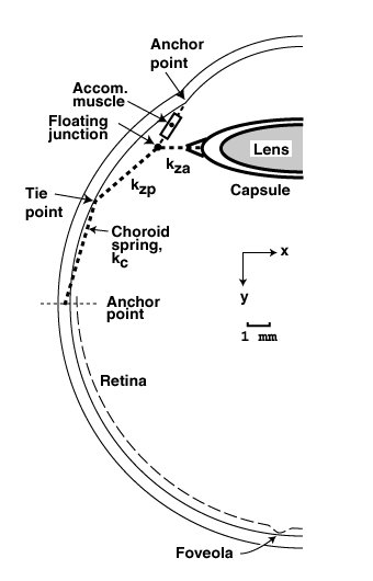
Note the floating junction connecting the accommodation muscle, the lens capsule, and the choroid spring. Note also the choroid spring consists of two sections, one labeled kc and one labeled kzp to conform to the curvature of the eyeball. Normally, when the eye is relaxed, it is focussed at distant objects. Under this condition, the accommodation muscle is relaxed and the choroid spring pulls the floating junction both outward (-x) and in the direction of the retina (+y). This action has two results. The link, kza is under significant stress and attempts to stretch the capsule to a greater diameter. This stretching tends to squeeze the lens to a thinner shape, thereby reducing the optical power of the lens. Simultaneously, the lens capsule is moved marginally closer to the retina to maintain optimum focus. [ It is important to note the eye is an "immersed optical system," the lens of the eye is a "thick" lens in optical theory, and its exit pupil (N') is not at the center of the lens.]
If an object of interest is at a lesser distance from the eye, the neural system senses this out-of-focus condition and causes the accommodation muscle to contract. When the accommodation muscle contracts, it has two actions. It pulls against the choroid spring thereby reducing the stress on the link kza. The edge of the capsule moves inward (+x) and the lens is allowed to assume a more spherical shape due to its own internal forces. This more spherical shape leads to a higher optical power lens and a change in both the (back) focal length of the optical system and the position of the exit pupil. Simultaneously, the lens capsule moves marginally in the -y direction to maintain optimum focus at the retina.
Following surgery, the accommodation system is essentially intact. However, the lens is now much more rigid and it is not subject to changes in shape or optical power due to the stress delivered by the link, kza. The radial position of the floating junction is now essentially fixed. However, the axial position of the floating junction remain subject to the activity of the accommodation muscle. Thus some axial motion of the lens is still possible under neurological control.
In the authors case, the residual error in optical power is accommodated by spectacles of small optical power when looking at distant objects. As a result, the accommodation muscle is essentially relaxed under this condition. When the spectacles are removed while still looking at distant objects, the neural system senses the out-of-focus condition at the retina and instructs the accommodation muscle to seek an optimum focal condition. Some neurological accommodation is/was perceived under this situation, particularly during the first few months following surgery.
The perception of post surgical accommodation may have decreased in recent times due to the natural reprogramming of the stage 6 memory associated with accommodation control.
This page is in beta release. The author welcomes and will respond to any comments or suggestions left at the comment page. Section numbers of the main manuscript, available on the web, are shown in brackets. The manuscript can provide more detail when desired. The first number shown is the chapter number; it is followed by the section numbers. Download individual chapters.
1. Beers, A. & Van der Heijde, G. (1994) In vivo Determination of the Biomechanical Properties of the Component elements of the Accommodation Mechanism Vision Res vol 34(21), pp 2897-2905
2