PROCESSES IN BIOLOGICAL VISION has defined a large and largely new set of performance descriptors applicable to the visual process in all animals. Some of these descriptors provide a new foundation for many of the previously defined empirical descriptors. It is important to rely upon a consistent set of terms in discussing these descriptors. The following list has been prepared to support the oculomotor generated motions of the eyes. The list is drawn from Section 7.3 of the complete text. It is also presented in support of the web pages related to the reading facility of humans. Further information and references concerning the operation of the oculomotor system of vision can be found in Chapter 7 of the text.
The oculomotor muscles of the human eye are known to be special, if not unique. They exhibit two distinct responses that are related to their morphology. The tonal part of each muscle is designed to act as a low frequency mechanical filter. That part exhibits a low frequency filter characteristic with a half amplitude point below 5 HZ. in response to an activation signal, consisting of a string of individual action potentials with a typical pulse width of less than 3 milliseconds.
A second, "twitch" part of each muscle responds entirely differently. It does not exhibit any filter characteristic and is known to "reproduce" the individual action potential waveforms. It is well documented that they can respond at frequencies up to 150 Hz or higher in response to individual action potential pulses of less than 3 milliseconds.
When the oculomotor muscles are combined in pairs, in order to drive the eyes either up-and-down or back-and-forth, they operate in a bang-bang mode to control the angular direction of the eyeballs. Many investigators have described the resulting motion as ballistic. Technically, the motion is not ballistic, particularly for the easily observable major saccadic motion. The more appropriate label is parabolic because the low frequency characteristic of the tonal muscles do not impart an impulse to the eyes, either when starting or stopping a saccade.
The oculomotor muscles are part of a very sophisticated servomechanism defined as the Precision Optical Servo System (POS). The neural portion of this system is centered on the pretectum of the midbrain. The pretectum controls the mechanical pointing of the eyes through a multimode sampled-data servomechanism. To accomplish this function, the pretectum extracts error signals, between the actual pointing angle and the desired pointing angle from the actual visual signals acquired by the retina. The system does not require or use any so-called efferescent copy of the neural commands issued by the pretectum.
The retinas of the higher chordates exhibit a specialized area of the retina that is known as the fovea based on its morphology. The fovea is generally circular and has a diameter of about 6.2 degrees in humans. Within the fovea is an even more specialized area based on its physiology. This inner fovea, or foveola, is the key to the operation of the analytical processes by which man evaluates images of objects occurring within the full field of view of his eyes. The foveola has a diameter, refered to object space, of only about 1.2 degrees. It is the limited extent of the foveola that demands the eyes of humans be highly mobile and be able to sweep the eyes through a large angle.
While many chordates (particularly the great hunting mammals and hunting birds) have foveas, and some show signs of a foveola, the capability has evolved to its peak in humans. This peak is directly associated with the remarkable sphericity of the human eyeballs.
As a result of the above capabilities, the motions of the eyes in response to the nervous system are extremely varied in amplitude and frequency spectrum.
The motions associated with the eyes of humans can be divided into two distinct groups: the major saccades that are easily observed by clinicians and researchers and the minor saccades that are virtually unobservable without special and demanding instrumentation.
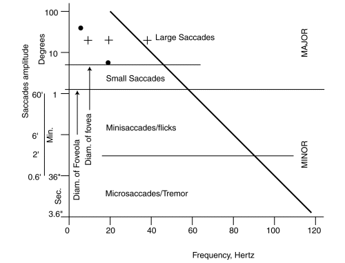
The major saccades are generally larger than 1.2 degrees and associated with the low frequency, less than 5 Hz, tonal response of the oculomotor muscles. They can be subdivided into the large, >6.2 degrees, and small saccades, between 1.2 and 6.2 degrees. The actual frequency response of the oculomotor system for major saccades exceeds 5 Hz because of overdriving of the muscles by the neural signals.
The minor saccades can also be subdivided into the minisaccades (or flicks) that are generally between 2 minutes of arc (0.033 degrees) and 1.2 degrees in amplitude and the even smaller microsaccades with a typical amplitude of 40 seconds of arc (0.01 degrees). The micro and minisaccades are generally associated with the twitch muscles of the oculomotor muscles. These muscles respond to much higher frequencies, between 40 and 150 Hz.
Figure 7.3.1-1 illustrates the above concepts.

It is difficult to establish the location of the slanting line in this figure. It depends on how some complex measurements are interpreted. The pair of black dots represent an alternate location based on one investigators measurements. The crosses show the measurements of another investigator over a period of years representing the same 20 degree rotation of the line of sight.
The twitch muscles are driven by a neural signal that can be described as a 90 Hz carrier modulated by a complex waveform associated with the scanning function. The modulations applied to the 90 Hz carrier signals applied to drive the lateral and vertical rectus muscles are virtually unrelated. The actual form of these waveforms is presented in Chapter 7 of the text.
As in the angular size of the saccadic motions, there is also a large range in the times required to accomplish these motions. This range can also be divided into those motions associated with general awareness, the analyses of specific objects and more specific analytical tasks such as reading, sewing and other craft skills.
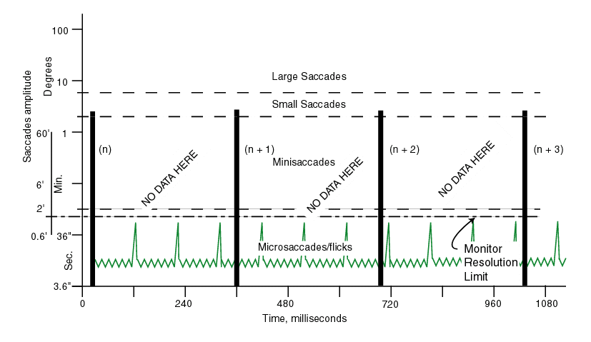
Large saccades occur relatively infrequently (typically in the 3-20 second range)and are frequently associated with changing the orientation of the head and body. Small saccades occur more frequently and have been the subject of most investigations into the mechanisms of scene perception and interpretation. Figure 7.5.1-2 shows a typical time line for the small saccades on viewing a new scene. Note the long intervals in which no data was recorded in these experiments. This was caused by the limitations of both the display device and the instrumentation. During these intervals representing a gaze, but frequently labeled with the term fixation interval, the eyes were not stationary or "fixed". They were performing the more analytical task of scanning a specific object for purposes of perception and interpretation. The minor saccades consist of the very low amplitude microsaccades associated with object or character group scanning and the slightly larger, but less frequent, minisaccades. The minisaccades or flicks are associated with changing the line of sight to view other features of the same object or the transition to the next character group in reading. These additional motions are shown by the colored lines in the figure. The time duration of the minor saccades are typically less than 10 milliseconds for the microsaccades and on the order of 10-20 milliseconds for the flicks.

The minor saccadic motions of the eyes, microsaccades and minisaccades (flicks), are the key to the ability of the visual system to perceive and interprete images. However, before discussing this subject, the reader should be familiar with the overall organization and mechanisms of the overall VISION facility, the operation of the READING facility, and the fundamental nature of the NEURONS.
With the above background in hand, the reader is directed to the DYNAMICS HOME PAGE for a discussion of how the crucial facility of feature perception and interpretation is implemented.