
Figures on this page are presented at reduced scale (resolution)to accommodate a browser. A larger scale version is available in Chapters 16 and 17 in the Download Files area that can be reached from the Site navigation bar.
All figures in this document are based on equal photon flux density per unit wavelength and not on equal energy per unit wavelength.
It has been conventional to consider humans as trichromats since Young in 1802. However, his proposal was only conceptual. He had no instrumentation that could prove his assertion. There is now excellent data showing the architecture of the human visual system uses and supports photoreceptors containing four distinctly different spectral absorbers (chromophores). This is the prototypical condition available in all animals (all biological visual systems). The architecture is presented in Section 16.2. This complete capability may not be expressed in all animals. However, there is a growing library of documentation showing it is present in many animals of all phyla.
The actual chromophores of vision are the Rhodonines. Their spectra are presented here. The complete discussion of the Rhodonies is in Chapter 5. The absorption spectra of the Rhodonines lead directly to the observed spectral responses measured by the psychophysicist and described on this page.
This page will address two performance area impacted by the tetrachromatic architecture of Human vision:
Until 1971, there had been no laboratory measurements of the spectral response of the human visual system starting at the retina. It was generally assumed that the performance of all elements of the visual system terminated near 400 nm. With the advent of modern surgical techniques, it has become common to remove damaged lenses from the human eye in order to improve overall vision. This damage may be due to trauma or to cataracts. The subjects of these operations have reported unusual sensitivity to very short wavelength light.
Virtually all previous tests on human vision in-vivo had included the physiological optics associated with the eye. Recently, psychophysical experiments have been performed on aphakic humans. Tan reported, and Griswold & Stark confirmed, that the spectral response of aphakic humans extended well into the area of 300-400 nm. Their data are shown in the following figure along with the theoretical spectrum developed in this work.
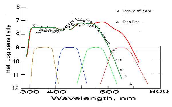
This figure extends down to below 300 nm unlike the conventional figures that stop at 400 nm or higher. Griswold & Stark provided the data represented by circles. Tan provided the data represented by triangles. The data points are within a range of 3:1. The solid curves are the theoretical overall spectral sensitivity functions (frequently labeled luminous efficiency functions) predicted by this work. The green line represents the predicted scotopic spectral sensitivity function. The red line represents the predicted photopic spectral sensitivity function. Details regarding this data and the theory are available in Section 17.2.2.
The absorption spectra of the individual absorbers within the photoreceptors of vision based on this work are shown at the bottom of the figure. They are shown normalized to a common value. These are the absorption spectra of the four chromophores found throughout the animal kingdom. See Chapter 5.
The theoretical spectra agree very well with the data. In general, the curves are between the two sets of data points. By adjusting the parameters of the theoretical function, either of the two sets of data can be fit even closer. The major deviation is at wavelengths beyond 600 nm. This is because of a problem in the experimental protocol. The subject was not operating completely within the scotopic range. The data points are beginning to transition up toward the photopic curve.
Griswold & Stark also measured the spectral response of the subject's other eye in order to quantify the absorption characteristic of the lens alone. They presented this data in the following figure.
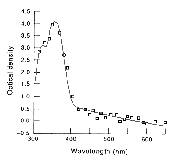
The solid curve in this figure is the calculated absorption spectrum of the optics. It consists of two parts. The left-most part is due to the absorption by alcohol and aldehyde radicals found in the material forming the lens. The long tail on the right is a portion of the second part. This part is due to Rayleigh scattering due to inclusions within the lens. These occlusions are due to the multilayer structure of the lens. The discussion supporting this figure are also found in Sections 16.3.3 & 17.2.2 The absorption of the macular is not included in the data or the theoretical curve.
Note that the above optical density is for a human lens of about 5 mm thickness. The optical density is proportional to the thickness of the lens. As will become apparent below, smaller animals have better ultraviolet visibility than humans because of their thinner lens. Larger animals have even less sensitivity in the ultraviolet and even blue regions for the same reason.
The following composite figure combines the absorption of the physiological optics and the spectral sensitivity of the human system starting at the retina. This figure is discussed in detail in Section 17.2.2.3 and briefly on the absorption page. It is the spectral response of a human subject when an adequate source of illumination is used.
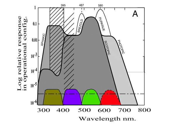
The hatching within the box illustrates the absorption of the normal lens. Without the lens the aphakic performance curve is obtained. With the lens, the senstivity indicated by the lower solid line is obtained.
To obtain these spectra in the laboratory, it is necessary that the source have a color temperature exceeding 6500 Kelvin (preferably 8000 Kelvin). It also requires the use of quartz envelopes for the light source and all external optics. With poorer instrumentation, the spectral performance of the eye is degraded at short wavelengths. This degradation is suggested by the hatching outside of the solid box. The degradation in this area has long been a source of debate in the empirical community. It was originally raised when Judd criticized the results of the C.I.E. Committee that defined the photopic luminousity function (and that he headed at the time).
The current literature has long had a difficulty describing the chromatic performance of the human eyes in the short wavelength region between 400 and437 nm. This region is hardly represented in the C.I.E. (1931) Chromaticity Diagram. Although it is frequently claimed that any color can be matched by a mixture of Blue, Green and Red. This is not true of the deep purple found near 410-420 nms. To match this color requires one of the matching lights to have a wavelength shorter than 410 nm.
The presence of four spectrally sensitive channels in the architecture of the human visual system (even though one is partly blocked on a wavelength selective basis) is very important in research. Besides expanding the number of photoreceptor (cone) types in the Young-Helmholtz Theory, it expands the number of chromatic axes in the Hering Theory to three. Because of this expansion, a three dimensional color space is required to describe human color vision completely (without introducing the black-white axes). Such a color space is presented below.
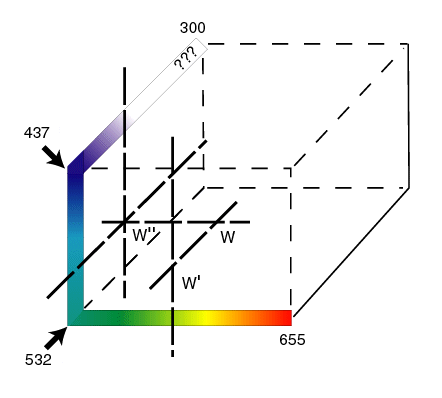
The derivation of this color space is presented in Section 17.3. Spectral colors with a wavelength between 300 and 437 nm are represented along the third axes drawn in the upper right. True purple occurs along this axis between 410 and 420 nm. There is a chromatic null along the third axis near 395 nm. The color at this point is defined as lilac. It plays a role similar to azure at 494 nm and "spectral yellow" at 572 nm. Within this color space, white is defined by the intersection of lilac, azure and spectral yellow. Thus white is described by three coordinates instead of the conventional two. The precise sensation of color perceived by the human visual system in the region from 300 nm to 395 nm has not been quantified.
A less cumbersome presentation can be obtained by rotating the upper axis into the plane of the conventional axes. There are two choices. First, a horizontal extension to the left as shown. While producing a graph that is not completely conformal, it does allow the colors that are mixtures of purple and blue through purple and green to be correctly represented in the second quadrant. Combinations of purple and red are not properly represented. Second, a vertical extension upward from 437 nm. This representation places purple above blue along the vertical axis. It represents combinations of purple and red but is unable to represent combinations of purple and green properly.
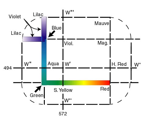
The rotation of the third axis into the vertical alignment results in a two-dimensional presentation that is quite useful except when trying to illustrate mixtures of purple and green. The resulting New Chromaticity Diagram for Research is available on this site and in Section 17.3.3.
As suggested above, if the light source is inadequate, the performance of the human visual system is degraded in the purple area. That is why artists and museums like natural skylight (not sunlight). When the system is degraded by inadequate illumination, a color temperature less than 7053 Kelvin, the performance of the human system begins todegrade in the purple area. Further degradation causes a loss in the perception of blues.
The architecture of the human visual system, along with that of most animals, is tetrachromatic. The performance of the human system at very short wavelengths is blocked by the absorption of its own optics. Therefore it can be more properly described as a blocked tetrachromat instead of a trichromat. The difference is significant in research.