
There is an architecture more fundamental to the neural system than the neuron itself. This architecture is illustrated below. The neural system consists of a large array of unidirectional signaling channels. These channels may branch and combine. However, the basic architecture is very simple.
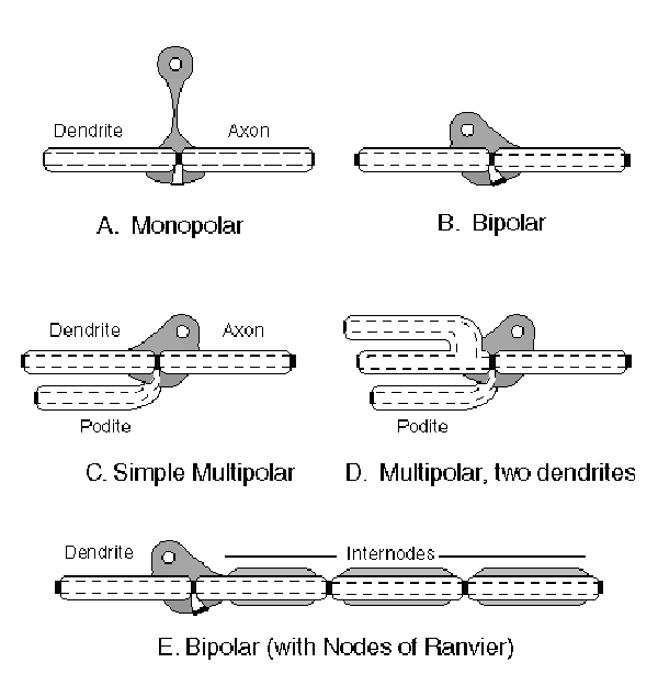
Fundamental types of neurons showing their internal signaling architecture
[from Section 8.4]
The fundamental signal path in the neural system consists of a series of electrolytic conduits connecting active electrolytic devices that can be considered as signal amplifiers (in the simplest connotation of that word). In this figure, the rectangular black bars at the end of and between conduit sections represent these active electrolytic devices, known as Activa ™. These devices are seen to appear in a variety of locations within and adjacent to the morphology of different neuron types. It is important to note that the devices between neuron types are found in morphological structures known as synapses while those found within a neuron have previously been unnamed (except for the hybrid situation known morphologically as the Node of Ranvier).
In A, the basic "monopolar neuron" is shown. This name is an archaic one. It was defined in terms of the number of "arms" emanating from the part of the cell containing the nucleus. As seen here, the nucleus plays no role in the neurological function of the cell. It is only concerned with metabolism and growth. As seen by comparing the "bipolar neuron of B with the monopolar neuron in A, there is no functional difference between them. The difference is one of convenience in morphological packaging (e. g, topographical arrangement).
The more important feature to notice is the presence of a third electrical path between the embedded Activa and the external surface of the neural cell. This third path has been named the podite. As in the case of the Activa itself, the nominal neuron is a three terminal device, not a two terminal structure as previously assumed in the literature.
When an Activa is found within a neural cell, the conduit associated with the base of the Activa is called the podite, in analogy with the dendrite. These two conduits comprise the neurites of the neuron and are generally associated with the signal input structure of the neuron. The axon is the third conduit and is normally associated with the output of the neuron.
In the monopolar and bipolar neurons, the podite is used primarily as a simple connection with the electrolyte surrounding the neuron. C and D show more complex neurons where the poditic structure has arborized and is used as a more typical input structure. These two frames only hint at the complex arborization possible in a single neuron.
It will be shown later that any change in input voltage applied to the podite will appear as an inverted signal at the axon. This is a crucial difference between a dendrite and a podite terminal.
E shows an important modification to a simple bipolar neuron, the incorporation of additional amplifiers (known as Nodes of Ranvier) within a single morphologically defined neuron. It is clear in this figure that the nucleus and other metabolic mechanisms of the cell are incidental to the primary signaling function. It is also clear that a so-called internode (or interaxon) acts as a morphological dendrite at one junction and as a morphological axon at a second junction. At the functional level, the internode is just an electrolytic conduit that exhibits a graded plasma potential between its two ends. The result is an optimized voltage between the plasma at each end and the adjacent podite terminal.
The above features are discussed in detail in Chapter 8.