
The following figures are presented here at reduced scale (resolution)to accommodate a browser. A version offering greater precision is available in Chapter 17 in the Download Files area reached from the Site navigation bar.
It is a little known fact that the human eye has a significant capability in the ultraviolet spectrum.. The complete spectral sensitivity of the human eye is shown in the following figure under a variety of conditions.
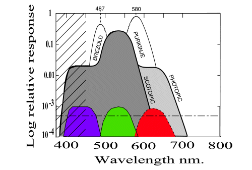
Variations in spectral response under various conditions [from Section 17.2.2]
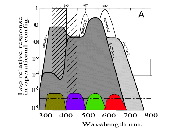
The spectral absorption characteristics of the four chromophores of vision are shown normallized at the bottom of the figure. These spectra are summed logarithmically to give the overall spectral sensitivity function of the retina. The darkest area is the region generally described as the scotopic spectral sensitivity function or frequently the "scotopic luminous efficiency function." The heavy line enclosing the scotopic area and the lighter gray area on the right defines the photopic spectral sensitivity function or the "photopic luminous efficiency function. The heavy line also enclosing the lighter gray area on the left defines the complete spectral sensitivity of the retina. This spectra is that perceived by a person who has had the lens of the eye removed (an aphakic eye. The heavy hatching represents the absorption of the lens. This absorption has a peak density of about 4 units at 357 nm and effectively nulls a majority of the sensitivity in the ultraviolet. However, even the normal eye has a significant sensitivity in the ultraviolet near 300 nm. The lighter hatching represents the loss in sensitivity, primarily in the 400-500 nm region due to Rayleigh scattering within the lens and other fluids of the physiological optical system.
Also shown are two areas of enhanced sensitivity under certain conditions due to the logarithmic summation process. These are generally known as the Bresold-Brucke Effect and the Purkinje Effect and occur near 487 and 580 nm. There is a potential equivalent effect near 395 nm in the aphakic eye.
The common wisdom has been that the human eye has no ultraviolet sensitivity and that its response to ultraviolet light was due to fluorescence within the eye. This is clearly not the case.
The scientific literature has only discussed and defined the spectral response of the human eye at wavelengths beyond 400 nm until very recently.
The luminous efficiency function of human vision varies considerably subject to the variations in source illumination and adaptation state as shown below.
Variations in spectral response at wavelengths greater than 400 nm under various conditions [from Section 17.2.2]
Under photopic conditions, the actual spectral performance varies significantly from the smoothed function V(l ) adopted by the CIE for a "Standard Observor." Although not often noted in the literature, the Standard Observor is neither a real observor or an average human observor. The accuracy of V(l ) has often been criticized, even by the Chairman of the Committee that adopted it. These variations will be discussed in abbreviated form below.
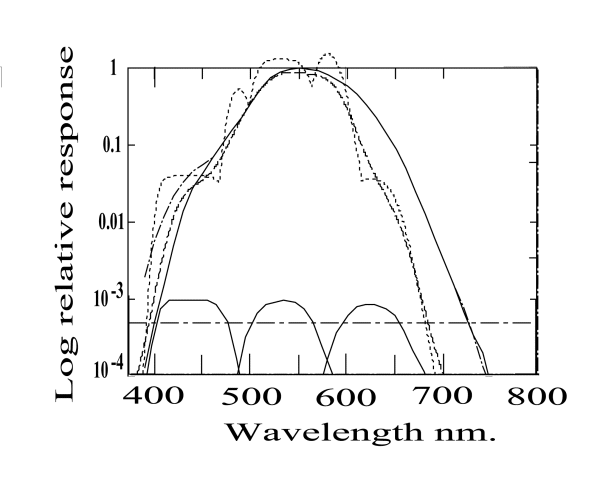
Spectral Response of the Human Visual System under photopic conditions
[from Section 17.2.2]
The theoretical perceived (as reported) spectral response of the visual system is given by the luminance channel signal as received at the cortex. This signal is defined by the logarithmic summation of the signals from the individual spectrally selective photoreceptor channels of vision. It should be noted that a logarithmic summation is significantly different from a linear summation under large signal conditions.
The above figure includes the spectral response of the three spectral channels of human (long wavelength trichromatic) vision. These responses are shown on a relative amplitude scale in the lower portion of the figure. Also shown is a horizontal line representing the one-half amplitude ordinate for these spectra. Whereas these individual spectrums show only limited overlap when displayed on a relative amplitude basis, this is not the way they are used in vision. In vision, the M-channel signal is given an emphasis of between ten and sixteen to one over the S- and L-channels. When summed under this condition, the theoretical photopic luminous efficiency function appears as shown by the (dotted line).
Note the theoretical photopic luminous efficiency function exhibits five individual relative peaks. Three of these peaks occur at nearly the same wavelength as for the individual spectral responses. However, there are two additional peaks due to the logarithmic summation process. These peaks are well known in the literature as the Brezold-Brucke phenomenon and the Purkinje phenomenon. These phenomena are most obvious under different conditions. Under hyperopic conditions, the peaks near 490 and 580 nm are both associated with the Brezold-Brucke phenomenon. Under mesopic conditions, the peak near 490 nm is not normally observed. The peak near 580 nm is associated with the Purkinje phenomena under these conditions.
All five of the theoretical peaks in the photopic luminous efficiency function are easily observed with adequate instrumentation. However, the historical measurements leading to the current CIE Photopic Luminous Efficiency Standard (solid line) were acquired using instrumentation of the 1920's (with corrections suggested by Judd using instrumentation of the 1930's). These instruments had an instantaneous spectral bandwidth of not less than 20 nm. When the results from various experiments were averaged, the result exhibited an average spectral bandwidth for the instrumentation alone of between 20 and 30 nm. Such instrumentation is completely inadequate for observing spectrums exhibiting spectral variations over intervals of 5-10 nm.
An additional problem has to do with the sampling plan used by these experimenters. Being unaware of the detailed structure of the waveform they were attempting to measure, they invariably centered their measurements at wavelengths that were multiples of 10 nm. This binning convention had the effect of further suppressing details of the spectrum occurring over intervals of 5-10 nm.
During the 1920's in particular, the importance of the color temperature of the light used to measure the photopic luminosity function was not well understood. Most of the measurements were made using incandescent light sources with color temperatures in the 2000-3000 Kelvin range. This had the effect of suppressing the measured spectral response of the eye in the blue region of the spectrum. Theoretical discussion occasionally addressed this insufficiency under the assumption that the visual process used "energy detectors." In fact the photosensitive mechanisms of animal vision employ "photon detectors." The theoretical and observed performance differences between these two cases are significant .
If the above instrumentation problems are taken into account, filtered (smoothed) versions of the theoretical function are found to approximate the CIE Standard quite well. In the above figure, the dashed line shows the predicted CIE Standard based on this theory assuming a 7053 Kelvin (equal photon flux per unit wavelength) light source and a 30 nm bin width. Although this curve does not match the CIE curve, it does if the excitation is changed from an equal flux per unit wavelength to the equal energy per unit wavelength condition. This condition is illustrated by the dashed-dot curve, still for a 30 nm bin width. This curve matches the published CIE curve, the solid line, precisely except in the blue region. The overall dashed-dot curve actually matches Judd's suggested modification precisely. The difference between the Judd curve and the CIE curve also surfaces the use of sodium glass lamp bulbs in the early experiments. These bulbs exhibit higher absorption in the blue region of the spectrum than anticipated, or documented, by the experimenters.
Note the shift of peak spectral response for the same function when plotted on an equal-energy basis compared to an equal-flux basis. The equal-flux curve has a peak 10-20 nm to the left of the nominal (and often quoted) 555 nm peak of the equal-energy curve.
The above features are discussed in detail in Part One of Chapter 17.
Return to the website home page