This page is in beta release. The reason is simple, it is now known that the human visual system does make use of the Ultraviolet (UV) photoreceptors in the retina in a third chromatic channel described by a third chrominance channel,
O = logUV - LogS. The high absorption of the lens in the UV region precludes this spectral channel playing a significant role in the luminance channel performance of vision. This revelation introduces a discontinuity in the New Perception-based Chromaticity Diagram (as well as in the CIE 1931 Chromaticity Diagram) along the 437 nm locus. While the New Chromaticity Diagram is rectilinear, projections on the diagram are not possible across the 437 nm discontinuity. The author welcomes and will respond to any comments or suggestions left at the comment page.
- - - -
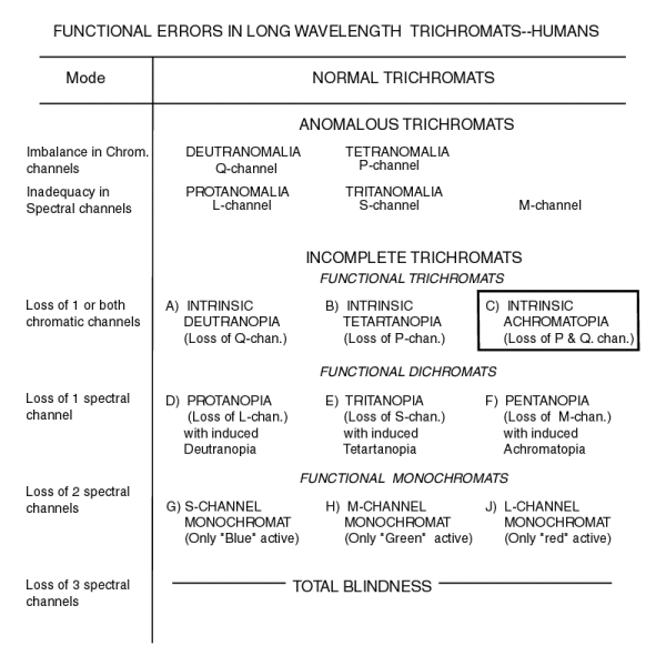
The literature is very contradictory concerning names for people with different types of abnormal color vision. The problem is primarily due to the ancient interest in the subject and the use of ambiguous generic names beginning in the 19th Century. This work attempts to use the generic names to the greatest extent possible to avoid confusion. This frequently requires the introduction of prefixes or modifying phrases as seen in the table below.
This table, from Chapter 18 of PROCESSES IN ANIMAL VISION, presents all of the possible types of abnormal color vision in a hierarchal form. It begins with the subject with normal color vision and ends with a totally blind subject. Between these two extremes are two major groups; those exhibiting anomalous properties and those exhibiting a total failure in one or more circuit elements of the visual system. The first are the anomalous trichromats and the second are the incomplete trichromats.

Historically a dichromat has been defined, based on observation of his performance, as a subject that could match any color using only two colors of variable intensity. Unfortunately, this is not an exclusive definition. All normal trichromats can match any color using only two colors of variable intensity. The two colors just need to be selected more carefully.
In this work, a dichromat is defined by the number of operational chromimance channels he possesses. A nominal trichromat has two functional chrominance channels. A dichromat has only one functional chromimance channel. These definitions say nothing about the number of functional spectral channels of the subject. However, as shown in the basic architecture of the eye, the loss of a spectral channel can have serious consequences. The loss of either the S- or L- spectral channel induces the loss of its respective chrominance channel and results in a dichromatic condition. The loss of the M- spectral channel induces the loss of both chrominance channels and results in a condition of achromatopia (total color blindness). Fortunately, the loss of the M-channel is unreported in the vision literature.
The loss of both chrominance channels without the loss of any spectral channels results in achromatopia without the loss of any spectral channels. This is the form of achromatopia suffered by the famous researcher, Knute Nordby. Nordby exhibited a normal luminance channel spectrum even though he was completely color-blind.
The above definitions clarify some older definitions. A deuteranope is a dichromat having lost his red-green discrimination capability due to the loss of performance in the Q- chrominance channel. A tetartanope is a dichromat having lost his yellow-violet discrimination capability due to the loss of performance in the P- chrominance channel. A protanope is an induced dichromat (a deuteranope) having lost his red-green discrimination capability due to the loss of the L- spectral channel (and the resulting loss of the Q- chrominance channel).
Using the new nomenclature above, it is possible to describe a subject with abnormal color vision much more precisely. Both unique New CHROMATICITY DIAGRAMS as well as old CIE (1931) Chromaticity Diagrams are presented for both the Deutranope and the Tetartanope. These figures answer the age old question: "What does a color blind person see?"
Also available are much more precise interpretations of readings taken with the Nagel Anomaloscope.