
As indicated earlier, the putative chemical concept of the synapse has never been extended to the description of the actual underlying mechanism or the resulting measured signal waveforms. Further, it has not even addressed the operating mechanism of the Activas found internal to the various neurons. The chemical concept must at least explain the operation of the putative neurotransmitters within the individual neurons as well as on their surfaces if it is to be creditable in this time period. The electronic concept in the literature has apparently gained adherents in recent years. In the electrolytic form proposed here, it is possible to describe the detailed mechanism associated with all of the active devices in the neural system, including those within individual neurons. This description includes the asymmetrical shape of some of these waveforms in unprecedented detail.
In the case of the Node of Ranvier as an example, all of the electrical characteristics associated with this feature can be provided based on the circuit model, shown in frame A above, when the Activa, shown as a rectangle, is replace with its symbolic equivalent. One of the results is the specific current paths and waveforms shown in the following figure.
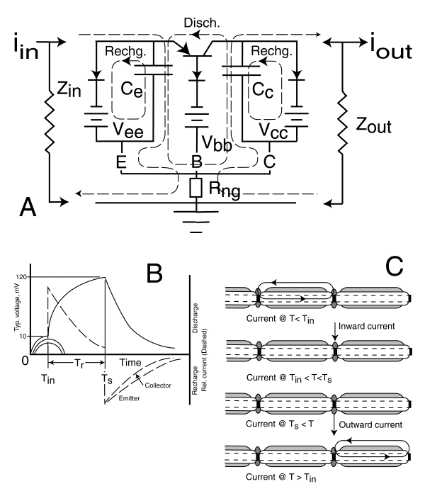
This figure is so complex, it cannot be explained in detail here. However, those details are discussed in the text. The Activa of a Node of Ranvier is operated in a switching mode. It only generates a single pulse, except when disturbed by pharmacological agents) in response to a stimuli. Frame A of the figure illustrates the various charging and discharging loops that occur as a result of this switching. It also shows the effective resistive impedance associated with the myelinated conduits connected to the Node. Frame B illustrates these individual currents as a function of time at a detailed level. These currents can be related to the currents shown in frame A of the previous figure. The important feature to note is the shape of the collector current shown by the solid line rising to a peak at time, TS, and then decreasing to the baseline. This waveform is clearly asymmetrical as seen in the more detailed waveforms found in the literature. In this respect, it is fundamentally different than the generator waveform of the photoreceptor cell. This waveform is an action potential. The waveform of the photoreceptor is not an action potential. This waveform also clearly defines the time delay, TR, associated with signal regeneration at a Node of Ranvier.
By overlaying these theoretical curves and delays on the published data, it is possible to determine the value of the circuit elements associated with a Node of Ranvier.
Frame C, in the lower right provides information in cartoon form concerning the signals at a given Node of Ranvier as well as the time of appearance of the same signal at the next Node. This information provides precise data concerning the capacitance and inductance per unit length of the conduits as well as allowing calculation of the loss factor in the conduit. This loss factor is related primarily to the dispersion of the signal within the electrolytic medium and is not the result of resistive dissipation of the energy of the waveform.
It should be noted that the loop currents shown in this figure are not in time synchronization with the currents marked inward and outward. This distinction is frequently overlooked in laboratory investigations where only the sum of these currents at a given time are measured.
Return to the NEURON Home Page.
Return to the website home page.