This animation describes the conversion of tha acoustic energy applied to the tympanic membrane into a surface acoustic wave on Hensen's stripe, a feature of the surface of the tectorial membrane leading to the Organ of Corti. This animation is unique in its explicit description of how energy applied to the oval window of the cochlea is actually processed on its way to the cochlea. No other explicit description of this phenomenon appears in the academic hearing literature. The source of information on the vestibule is the surgical literature.
The primary purpose of the auditory canal is to collect the energy provided by the external ear and create an acoustic wavefront that is parallel with the curvature of the tympanic membrane. This energy is used to move the tympanic membrane piston style, in and out. The extent of the motion is quite small. As a result, the tympanic membrane can be considered a solid wall when studying the initial wave motions within the auditory canal and the reflections thereto. On the other hand, the small motion of the tympanic membrane is levered by the ossicle bones into a larger hydraulic motion within the much smaller volume of the vestibule. The hydraulic energy is further concentrated into the still smaller cross section of Hensen's stripe. The resulting energy density within Hensen's stripe is orders of magnitude higher than that at the tympanic membrane.
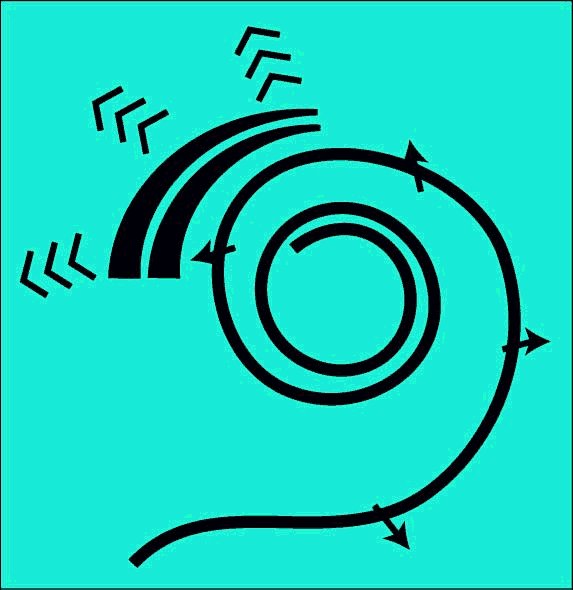
The animation begins with the application of four square-wave pressure pulses to the tympanic membrane at the end of the auditory canal. Each pulse causes a small motion of the membrane and the associated ossicle bones. The motion of the stapes moves the diaphram within the oval window back and forth. Each motion of the diaphram causes a longitudinal pressure wave to spread within the fluid of the vestibule as shown. As each pressure wave impinges upon the end of the cochlear duct extending into the vestibule (the vestibular caecum or cul-de-sac of the cochlear duct), the energy of that wave is converted into a surface acoustic wave traveling at nearly a right angle to the incident wave. This surface acoustic wave exits the vestibule on the underside of the cochlear duct and proceeds along the edge of the cochlear duct as shown. The microscopic structure at this edge is known as Hensen's stripe.
The pulses traveling along Hensen's stripe are high-quality reproductions of the incident square-waves. These pulses are analyzed for their frequency and temporal characteristics at the next stage of the system as shown in subsequent animations.
The fundamental task of the vestibule of the labyrinth is to provide an environment for converting the hydraulic energy applied to the fluid within the vestibule into a modified surface acoustic wave (Modified Rayleigh wave) traveling along Hensen's stripe within the cochlear partition found along the centerline of the curved cochlea immediately above the basilar membrane.
One unique characteristic of this energy conversion is the large change in propagation velocity of the energy. In the auditory canal, the pneumatic acoustic energy propagates as a longitudinal wave at 344 meters/sec in air. Within the vestible, the hydraulic acoustic energy propagates as a longitudinal wave at 1500 meters/sec. After conversion into a surface acoustic wave, the acoustic energy propagates at a transverse wave at only 6+ meters/sec (a reduction of 57:1 over the pneumatic velocity).
In the fluid environment of the cochlea, the propagation velocity of 1500 meters/sec means a 1000 Hz signal has a wavelength of 1.5 meters(60 inches). This wavelength is 43 times the uncoiled length of the cochlea. Such a disparity essentially eliminates the possibility of any distributed resonance phenomenon associated with the fluids of the cochlea. At the reduced propagation velocity of the surface acoustic wave (6+ meters/sec), the situation is quite different. The 1000 Hz signal has a wavelength of only 6+ millimeters (less than one fifth the length of the uncoiled cochlea). While no sign of any resonance phenomenon has ever been identified within the cochlea, this wavelength is compatible with a dispersion phenomenon.
At the slow propagation velocity of 6+ meters/sec, the delays associated with the acoustic signals are on the order of six milliseconds for a signal traveling the full 35 mm length of the uncoiled cochlea. This is the same value found in the empirical literature for signal of 60 to 100 Hz.
The unique convoluted triangular shape for the cochlea shown on the right was obtained by filling the scala media (the space between the Reisnner's membrane and the basilar membrane) with a plastic before striping away the structure forming the scala vestibuli and the basilar membrane. The ridge closest to the viewer indicates the location of the Organ of Corti and the tectorial membrane covering it. Note the scala vestibuli and the scala tympani play no active role in the hearing mechanism.
The underlying frequency dispersion mechanism, described in the next animation, requires a curved cochlea. A straight, or uncurled, cochlea is not a functional cochlea. While the cochlea need not be coiled as in the higher mammals, it must be curved. It is the local curvature of Hensen's stripe that provides the mechanism for frequency component separation.
The resulting surface acoustic wave moves along the ridge in the figure that corresponds to the location of Hensen's stripe and, at a larger scale, the Organ of Corti. Once the energy enters the spatial dispersion and frequency analysis section, the energy is dispersed as a function of frequency as seen in the next animation.
Within the region labeled the Launcher region, the curvature of the cochlear partition is in a direction that does not interfere with the propagation of the modified surface acoustic wave. In the frequency analysis region, the curvature is such as to disperse the energy in the surface acoustic wave as a function of frequency.
For the scope and top-level description of the SAW-based Electrolytic Theory of Hearing, see page 18 of Chapter 1 of the e-book "Processes of Biological Hearing"
The above material related to the conversion of the acoustic energy applied to the tympanic membrane into a modified surface acoustic wave (modified Rayleigh wave) is drawn from Section 4.2 of Chapter 4 of "Processes in Biological Hearing." A draft of this material can be accessed from the Home Page of this website.
Go to the next animation in the operational chain. XXX NOT ACTIVE YET
Go to the roadmap of available animation of hearing. XXX NOT ACTIVE YET
Return to the website home page.
Anson, B. & Donaldson, J. (1992) Surgical Anatomy of the Temporal Bone, 4th Ed. NY: Raven Press pp 71 & 267-277