
The precise method of acoustic energy extraction used in hearing has been a mystery for a very long time. The mystery has been nurtured by the very small size of the cochlea compared to the wavelength of acoustic waves in moost biolotical materials. There is a special material within the cochlear partition that has properties uniquely suited to the extraction of the acoustic energy applied to the cochlea. The material is a liquid-crystalline (or gelatinous) material coating the side of the tectorial membrane facing the auditory sensory neurons. These sensory neurons have been called "hair cells" for a very long time based on a broad translation of the Latin word cilia.
The basic mechanism associated with the above liquid-crystalline material involves a surface acoustic wave (SAW). Multiple discrete surface acoustic waves subsequently interact with the piezoelectric elements of the auditory hair cells.

The mechanism described further below is used in all mammalian, reptilian and avion hearing. The mechanism is used in the hearing of the monotremes, the egg-laying but nursing animals such as the platypus and the echidna of australia. A similar mechanism may or may not be used in amphibians.
For the scope and top-level description of the SAW-based Electrolytic Theory of Hearing, see page 18 of Chapter 1 of the e-book "Processes of Biological Hearing"
The following material related to the energy extraction mechanism within the cochlea is drawn from Section 4.3 of Chapter 4 of "Processes in Biological Hearing." A draft of this material can be accessed from the Home Page of this website.
The challenge faced by the cochlea is to accept acoustic energy into a fluid environment where the velocity of sound is typically 1500 meters/sec or more. In this environment, the wavelengths of the propagating energy are much greater than the size of the cochlea. The frequencies and wavelengths of interest in such a medium make their sensing by resonance based techniques extremely difficult. The challenge is to slow the velocity of propagation of the energy considerably and avoid the limitations of resonance based sensing techniques. This animation will focus on the first problem, the propagation of the energy. A later animation will concentrate on the dispersion technique used to side-step the resonance problem.
The gelatinous coating of the tectorial membrane is a liquid-crystal. It does not exhibit any structure at the anatomical or microscopic level. Its structure is at the molecular level. The structure, which is common to edible gelatin, consists of a super crystalline-like lattice within the liquid composing 99% of the bulk of the material.
Gels of the type described above are capable of supporting a special form of energy propagation at their surface with a material of lower density. It is a surface acoustic wave of the type first studied by Raleigh during the 19th Century and now colloquially called Rayleigh waves. A surface acoustic wave propagates only at an interface between two materials of different density. The velocity of propagation in the higher density material, the gel in this case, can be much slower (orders of magnitude slower) than in the other material.
Simple gels are widely known for their very low attenuation factors for acoustic energy. They are also known for their very low surface tension. These properties have led to a series of simple laboratory experiments to demonstrate the "ringing of gels." The frequency at which they ring is determined more by the container holding them and the method of excitation than by the properties of the gel itself. The low propagation velocity, low attenuation and low surface tension make them ideal for use in the cochlea.
The gel surface of the tectorial membrane supports the propagation of acoustic energy as a surface-acoustic-wave (SAW-wave), or Raleigh wave, traveling at a nominal 6+ meters/sec in humans. This is 250 times slower than a compression wave in a fluid. The surface acoustic wave is propagated without significant loss or group delay for frequencies between 10 and 150,000 Hertz in mammals.
The transition from a compression acoustic wave within the vestibule of the labyrinth to a SAW wave within the cochlea is accomplished as it is in many man-made devices, by a launcher. No Latin name has been associated with this structure previously. This element is made of the gel material but has a specific shape optimized for accepting a compression wave at one surface and transferring the energy to a surface acoustic wave on another surface. The energy then propagates along this second surface at a lower velocity than the incident compression wave. As will be shown in a later animation, the propagating energy is concentrated at the edge of the gel in a structure known as Hensen's stripe.
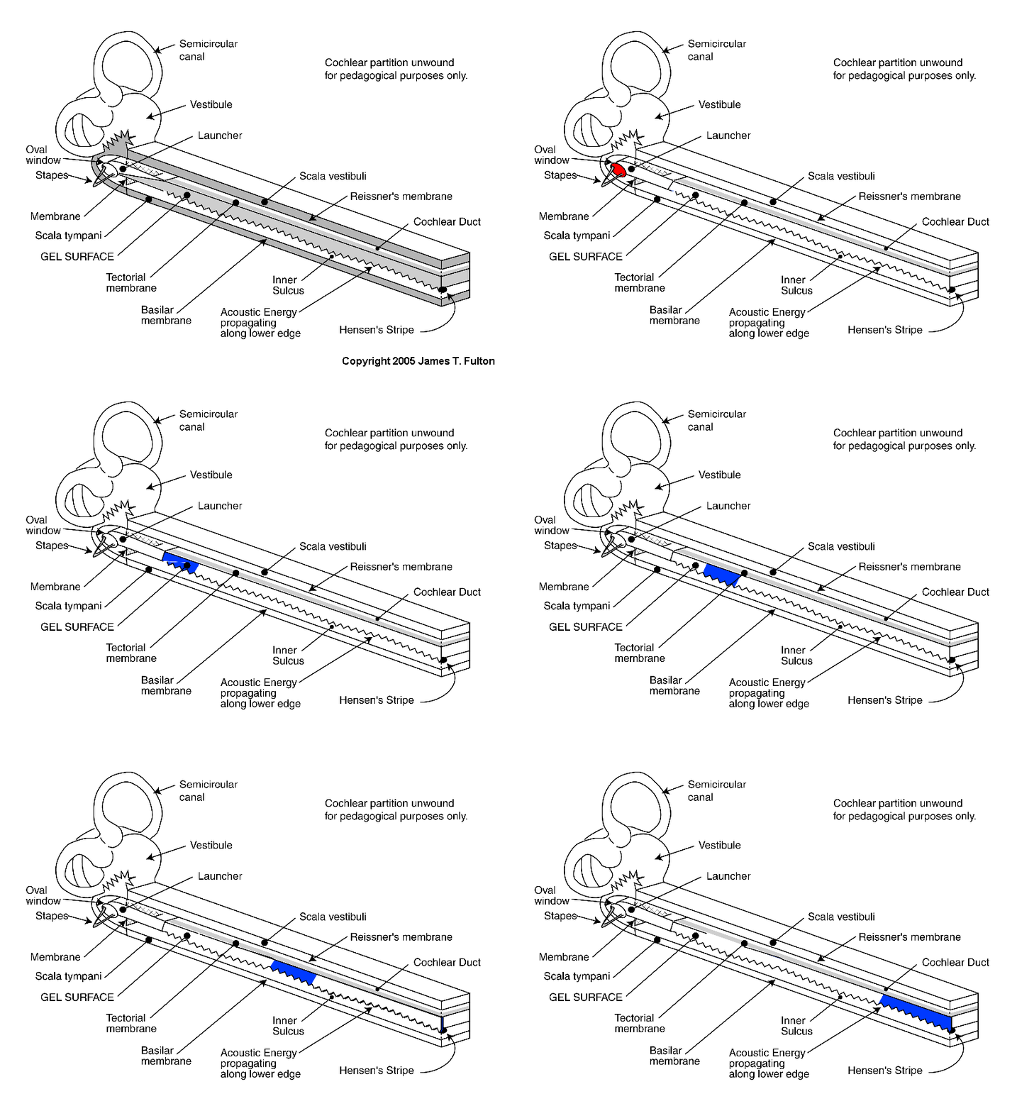
The following animation is meant to describe how a square pulse of energy applied to the oval window of the cochlea is processed within the cochlea. It is basically a two-step process. The energy is initially in the form of a fast-moving compression wave in a liquid environment. It is converted to a slow-moving surface acoustic (or Raleigh)wave that progresses slowly down the length of the gel surface within the uncoiled cochlear partition. It is important to note that the cochlea has been uncoiled for illustrative purposes only. The next step in cochlea operation, individual frequency component isolation, requires that it be curved.
The first frame of the animation is provided for orientation purposes. Callouts are provided to all of the relavent portions of the cochlea. Following this frame, an initial unshaded frame is introduced. Following this frame the animation sequence begins.
Initially, a square pulse of energy is applied to the oval window for a finite time (shown in red). This pulse propagates into the fluid chambers of the labyrinth where it is restricted by the small size of the paths leading away from the main vestibule. Only a small percentage of the energy can propagate into these areas (shown in orange). However, the high propagation velocity of this energy causes the pressure within the entire labyrinth (including the scala vestibuli and scala tympani) to equallize within a very short period of time (in less than 0.050 millisec.), much less than one cycle of most frequencies of interest in hearing. As a result, no significant differential pressure persists within the cochlea due to the propagation of the compression wave.
The majority of the initial energy (probably over 80%)is applied to the area occupied by the launcher. The launcher accepts the propagating compression wave energy (shown in red) and converts it into a slowly traveling surface acoustic wave (shown in blue)
The slowly propagating surface acoustic wave (shown in blue)moves down the gel surface at approximately 6+ meters/sec in humans. It travels as a transverse wave along the surface of the gel, as shown by the complex sine-wave, taking a total time of about 6 millisec.). For convenience, the energy of the surface acoustic wave is shown as a constant shade of blue as it moves along the gel surface of the tectorial membrane. In fact, energy is extracted from the packet as it progresses, beginning with the removal of the high frequency information first. Thus, if the shading was related to the total energy content, it should fade to a pale packet as it approaches the apex of the cochlea

Go to the next animation in the operational chain. XXX NOT ACTIVE YET
Go to the roadmap of available animation of hearing. XXX NOT ACTIVE YET
Return to the website home page.